Not every pink macule is a hemangioma-when a correct diagnosis is the basis of effective treatment. Vascular anomalies are a very frequent pathology in the daily practice of pediatricians. According to the vascular anomalies’ last classification (ISSVA classification revised in 2018), all vascular anomalies are divided into two main groups: vascular tumors and vascular malformations.

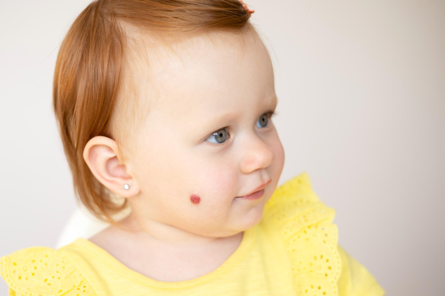
The most common vascular anomaly in children is Infantile hemangioma (IH). It is a benign vascular tumor that occurs in 3-10% of newborns.
It could appear everywhere on the body, but mostly on the head, neck regions, then trunk and extremities. IH has stages of its development: rapid proliferation, stabilization, and involution.
There are superficial, deep, and mixed infantile hemangiomas depending on which depth in the skin it is located. In the majority of cases, there is no need for treatment. But approximately 10% of IH, which are classified as high-risk IH, must be treated as soon as possible, ideally during the first months of life.

The high-risk IH are those which could lead to life-threatening complications, functional impairment, bleeding, ulceration, disfigurement, multiple hemangiomas (5 or more), and also those which are part of some syndromes.
When there is a suspicion of such syndromes, for example, PHACE syndrome and LUMBAR syndrome, there are some investigations (Doppler US, MRI, MRI with angiography, echocardiography, etc.) and consultations with other specialists, which must be done to confirm the diagnosis and further follow up. In these cases, the patient must be referred to a specialist as soon as possible, which will make a correct diagnosis, prescribe the proper treatment, and do further follow-up.
Nowadays, the first-line treatment of IH is propranolol, in a dosage of 2-3mg/kg divided into 2 doses. It has indications and contraindications. As mentioned earlier, propranolol is started as efficiently as it is. In severe cases, when a rapid effect is needed or cases when oral propranolol is contraindicated, systemic corticosteroids could be prescribed. In superficial IH, the topical timolol maleate 0.5% solution or gel is prescribed twice daily. A pulsed dye laser is used mostly for correcting the residual lesions.
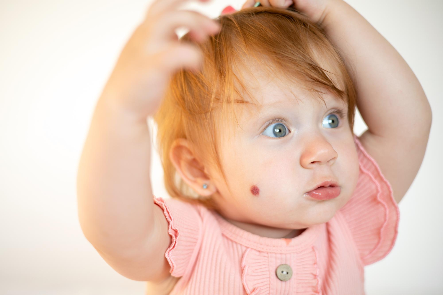
But very often, pediatricians wrongly diagnose every pink macule as a hemangioma. As mentioned above, there are also different vascular malformations, which could resemble IH at first impression.

In vascular malformations, there is a defect of some vessels but not a proliferation of vessels as in vascular tumors. In pediatric practice, the common vascular malformation is a capillary malformation, where there are dilated capillaries in the skin. The most common capillary malformation is Nevus simplex or salmon patch, which is seen in 30-40% of newborns. It is a benign lesion and, in most cases, resolves spontaneously before 2y old. There is no need for investigations or treatment.
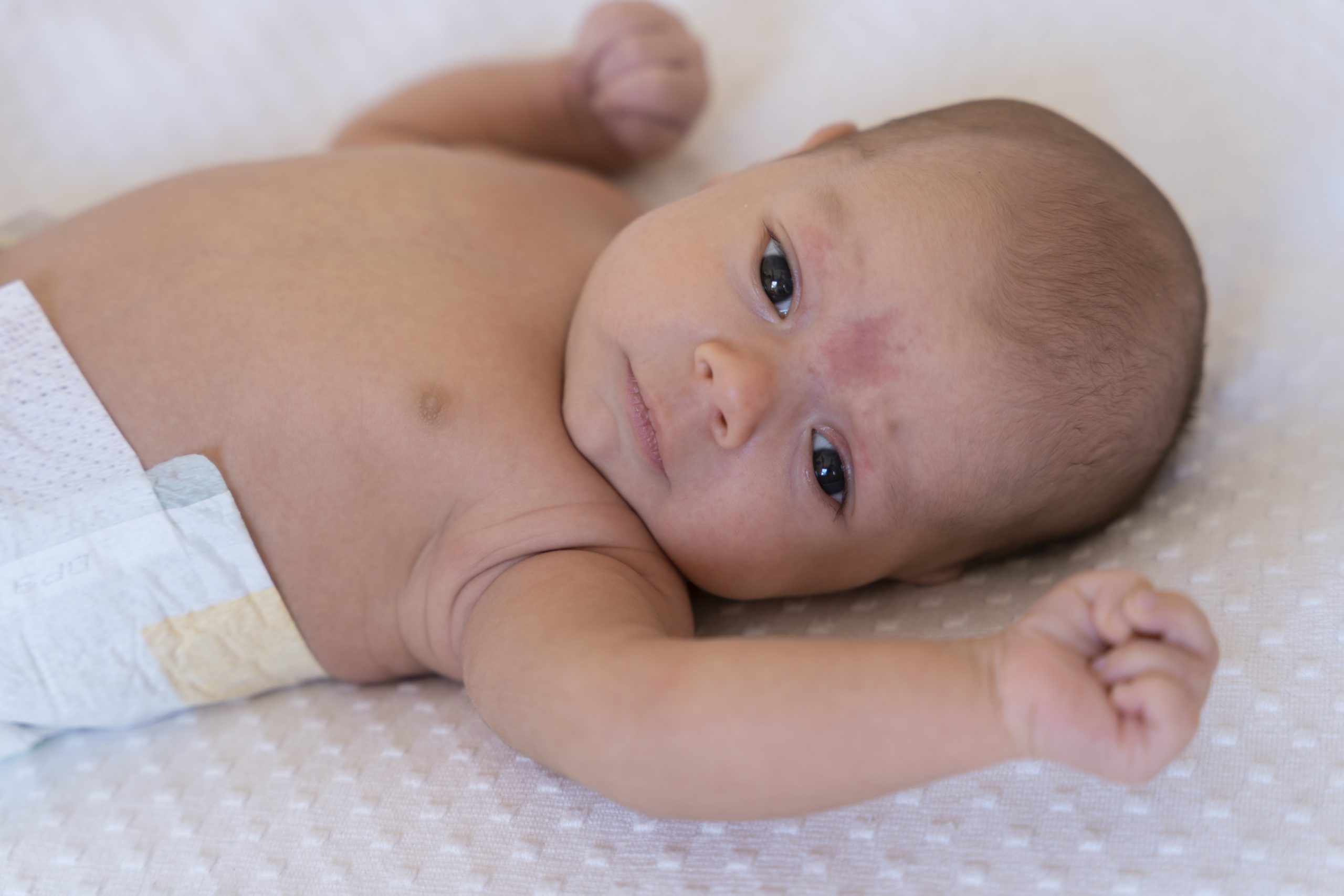
Nevus flammeus, or port-wine stain, is another capillary malformation. It occurs in 1/300 newborns. Usually located on the face, unilaterally. It does not disappear with age and even becomes darker and thicker. But besides its esthetic disfigurement, the port-wine stain could also be a sign of different syndromes, such as Sturge Weber, Klippel Trenaunay, or others, when the correct diagnosis and early intervention are mandatory.
it is very important to differentiate IH from vascular malformations or other hemangiomas because the approach and treatment are different in any case. For example, propranolol is not effective in Nevus flammeus, and we must avoid unnecessary prescriptions of this drug.
In conclusion, we must emphasize that all pediatricians must be aware of vascular anomalies to correctly diagnose infants. Depending on the diagnosis, the further approach could be different -active intervention or a follow-up.
In all cases, when the diagnosis is suspicious or when there is a high-risk IH, the patient must be referred to specialized centers. Arabkir joint medical center in Yerevan is one such center, where pediatric dermatologists, together with pediatricians and cardiologists, and radiologists, do teamwork, from diagnosis to follow-up and treatment, both inpatient and outpatient.
Hope this helps!
Best regards,
Adilkhanyan Armine, Head of the Pediatric Dermatology service, Arabkir JMC.