Usually, when speaking about melanocytic naevi in children, one needs to incorporate topics like congenital and acquired melanocytic nevi (AMN), as well as Spitz, blue, nevus spilus, and halo nevi. Here I am going to concentrate on acquired melanocytic and Spitz nevi.
Generally, acquired nevi (moles) in children are not a source of concern. Even though changes in the number, color, or size of nevi are typically observed during childhood, malignant neoplasms like melanoma are rarely diagnosed. Regardless of its rarity, the ability to raise suspicion about atypical lesions is crucial because melanoma is a highly treatable condition when diagnosed early.
Appearance and behavior of acquired melanocytic naevi in adults do not follow the same rules as those in childhood, especially in the first ten years of life and during puberty. Knowing several key aspects in this age group will help avoid anxiety and decide when a dermatologist should see the child.
It is always better to examine the whole child rather than one nevus because most nevi in an individual display phenotypic stability and look like one another. Nevi that diverge from the “signature” look is called “ugly ducklings” and need to be closely examined.
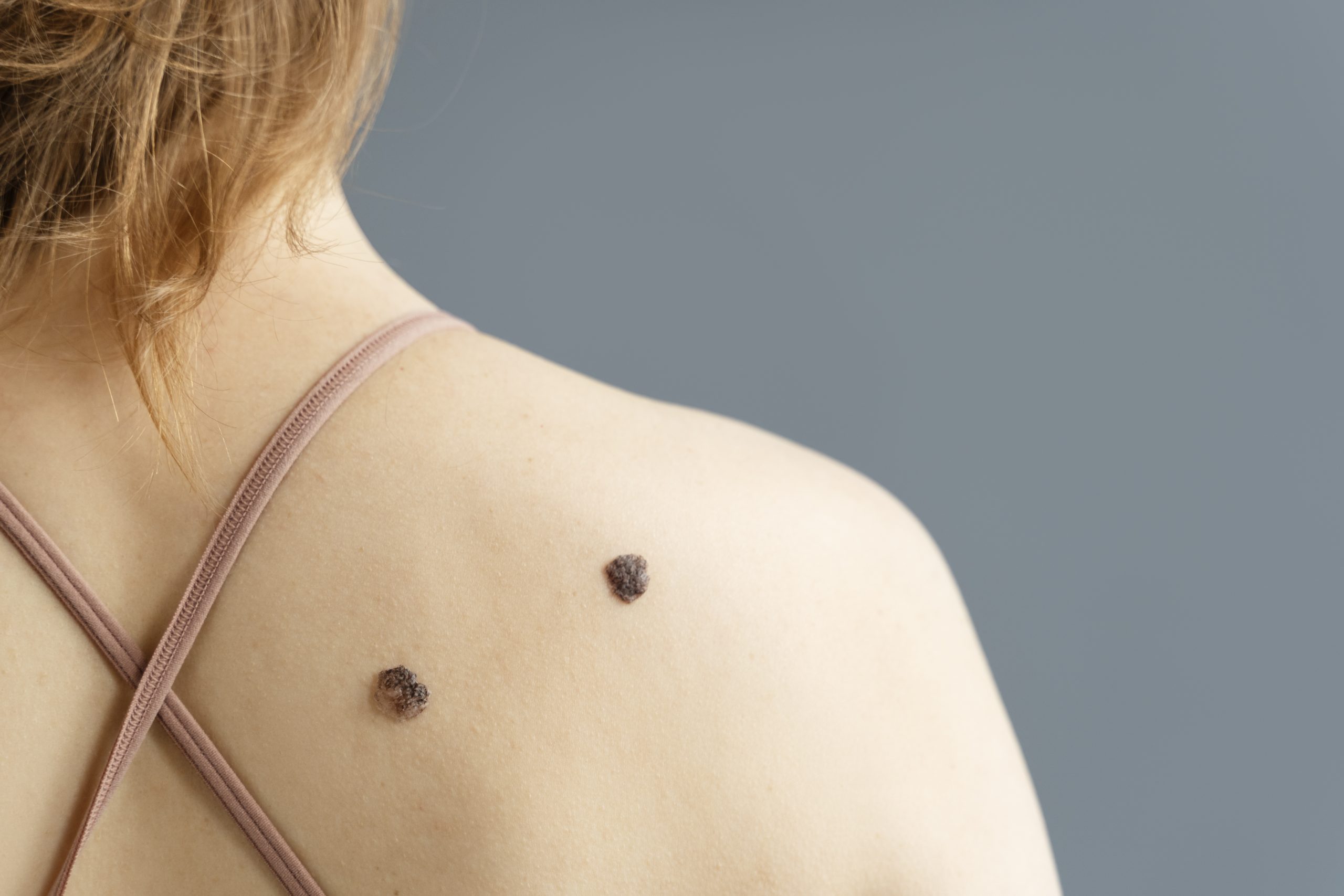
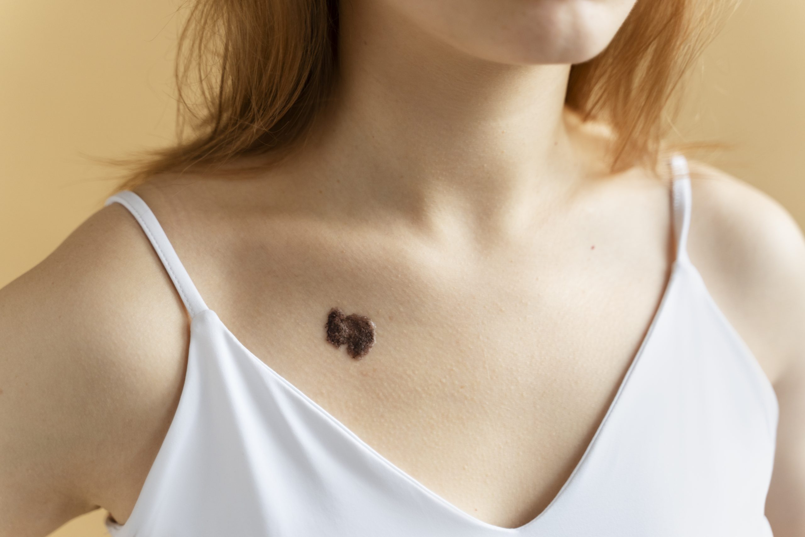
Typically acquired melanocytic nevi in childhood are 1-5 mm in diameter, and only a small portion may be bigger than that. However, growth in proportion with the child is commonly observed during puberty. Change of this type is not enough to raise suspicion of melanoma in this age group. AMN is usually brown to black in color and often may get darker or lighter during childhood. Multicolored lesions should also raise suspicion. In case of very rapid changes in size and color, dermatologists should be consulted.
AMN is generally symmetric, with a round or oval shape. Congenital nevi or nevi acquired very early in life usually do not follow this pattern.
The mean number of acquired nevi until becoming an adult is 20. If multiple nevi are present (more than 50), the child must be examined by a dermatologist.
Junctional naevi, or nevi in which nests of melanocytes are located in the epidermis, are not palpable. Nevi, in which melanocytic nests are located at the epidermal-dermal junction and the dermis (compound nevi) or the dermis (intradermal nevi), have a palpable, usually smooth surface. Sometimes this dome-shaped surface may get traumatized or irritated, but bleeding of the nevus or ulceration without a history of trauma may be a sign of melanoma.
Dome-shaped nevi that are pink, red, or red-brown may be Spitz nevi. These benign melanocytic nevi are usually a source of anxiety and are sometimes hard to differentiate from melanoma clinically and histologically. They usually appear between 10 and 20 years of age but also may be congenital. The dome-shaped surface may bleed and be unsmooth on palpation. A dermatologist should examine lesions that resemble a Spitz nevus. Dermoscopy is a particularly useful tool in the diagnosis of Spitz nevi.
The diagnosis of Spitz naevus is usually confirmed by an excisional biopsy. In children under the age of 12 years, a Spitz naevus may be monitored using dermatoscopic surveillance. The normal evolution of Spitz nevi is rapid growth for approximately six months, stabilization for a few years, and spontaneous resolution.
Hope this helps!
Best regards,
Dr. Lida Melikyan, Dermatologist of “Wigmore” Medical Centre