Background
Spinal deformities are one of the most common causes of referral to a pediatric orthopedic surgeon. There are many false stereotypes and ideas about these deformities that have been propagated for many years by non-specialists and in textbooks.
Varieties
There are a few subtypes of scoliosis (e.g., congenital, symptomatic, neuromuscular, and idiopathic). Congenital scoliosis manifests as irregularly formed vertebrae, while symptomatic scoliosis is usually the result of other musculoskeletal conditions. Some neuromuscular diseases, such as myopathy or cerebral palsy, can also cause spinal deformity, usually leading to big C-type deformities. There are subtypes of idiopathic scoliosis. It can be early onset, which starts before the pubertal spurt. This type is quite aggressive and causes severe and stubborn deformities, but fortunately is not very common. Another type is adolescent scoliosis, which is the most common form of scoliosis and is most prevalent in girls.
In instances where there is suspicion of an underlying spinal abnormality or abnormal neurological findings, obtaining diagnostic information is important. This is particularly relevant for subgroups of individuals with a higher likelihood of spinal abnormalities, such as those with left convex thoracic curvature or symptomatic scoliosis. In these cases, an MRI can provide valuable information for developing effective treatment plans.
Causes and Mechanisms
Although the cause of idiopathic scoliosis is unknown (that is why it is called «idiopathic» ICD10 M4.1), the mechanism of the disease is well known. Idiopathic adolescent scoliosis results from the disparity between the growth of the vertebral bodies both anteriorly and posteriorly, leading to the deviation of the entire spine.
Conservative Treatment
The treatment of the disease starts by considering the mechanism of deformation. As scoliosis appears, it is important to identify the remaining growth of the child. To assess it in individuals, radiographic signs can be used alongside other important diagnostic indicators. Information from the patient’s medical history, including secondary gender appearance, menstruation, and parents’ height, can provide clues to their current stage of development. Additionally, clues from a physical exam, such as the pubertal stage, can help determine the remaining growth potential. If the child is determined to still be growing, and the spinal curve angle is below 20 degrees, then only follow-ups are needed approximately every 4-6 months. During these follow-ups, the progress of the spinal curve is assessed clinically (by checking rib bump height and balance) and radiographically. If the angle exceeds 20 degrees, then bracing is the only approved treatment.
About Brace
The brace is quite a rigid device that is made from special plastic. It often includes the lumbar and thoracic parts of the patient. The child is growing 24 hours a day; thus, it is indicated to wear the brace for almost the same time period (22-23 hours a day) to influence growth. The patient uses the remaining time without the brace for hygienic procedures and sports activities.
It is important to note that the main goal of the brace is to prevent curve deterioration or to reduce progression at the very least. If it is possible to keep the curve under 40 degrees until skeletal maturity, the curve progression tends to halt, and there is no need for further treatment. After growth ends, the wearing of a brace is not indicated, as it is not effective.
As an alternative treatment for a brace, the doctor can suggest casting. Usually, it is indicated in elastic and big deformations as the first step before bracing to achieve better correction and improve the result of treatment.
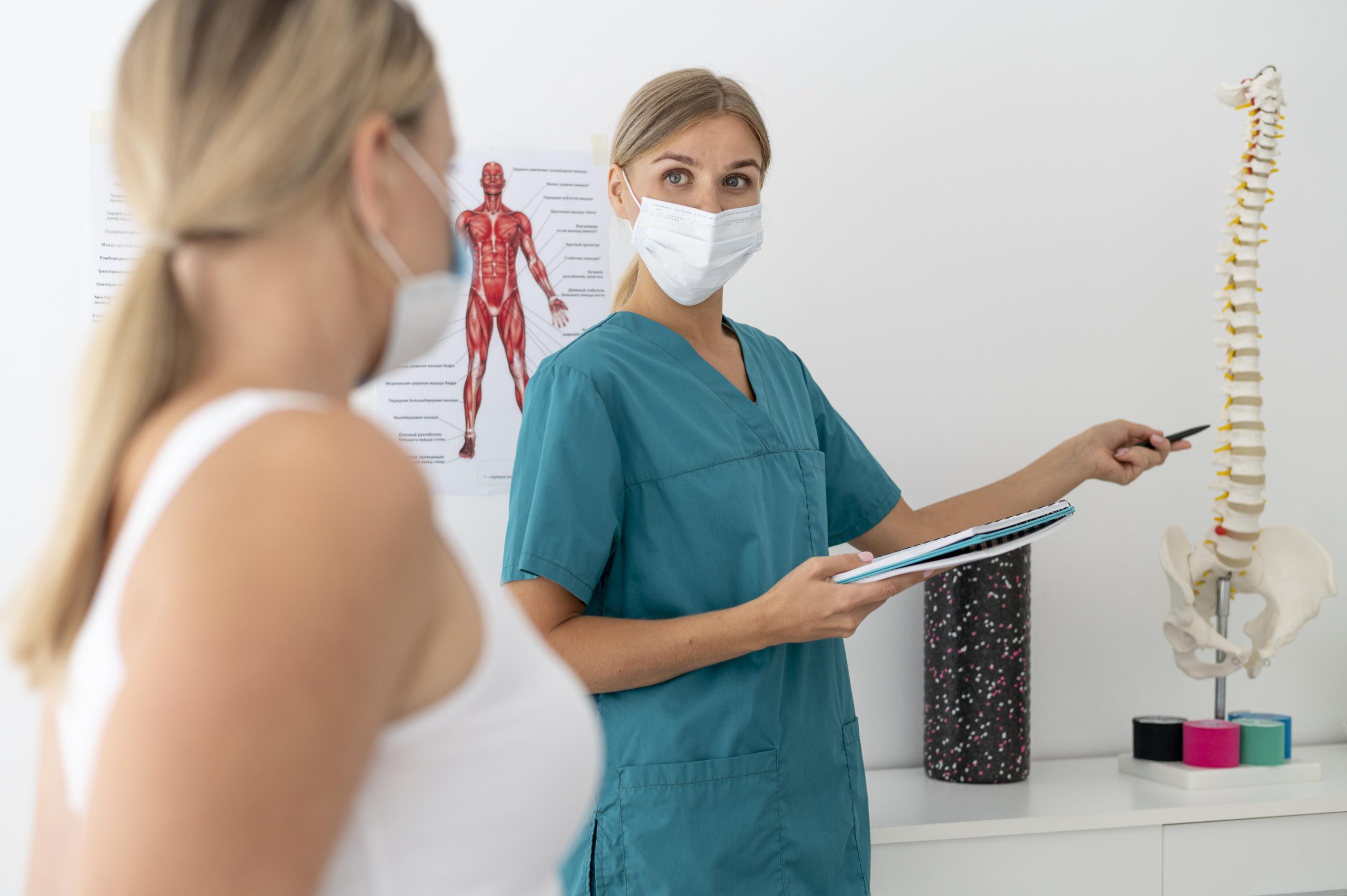
Operative Treatment
If the curve at skeletal maturity is more than 40 degrees, then the curve magnitude tends to continue progressing even if there is no growth remaining and operative treatment is required. It is possible to correct the curve during the operation of about half of the existing curve; however, the main goal is to stop the curve progression and balance posture in sagittal and frontal planes to have the head above the center of the pelvis. The cosmetic issues are secondary.
The operation involves placing screws into the vertebrae, which are then fixed to each other by metallic rods. After that, the deformed segment of the spine is corrected and immobilized.
Mobility of the spine does not impair after this operation, and the patient can return to his/her daily activities after the wound and surgical place are healed.
Misconceptions
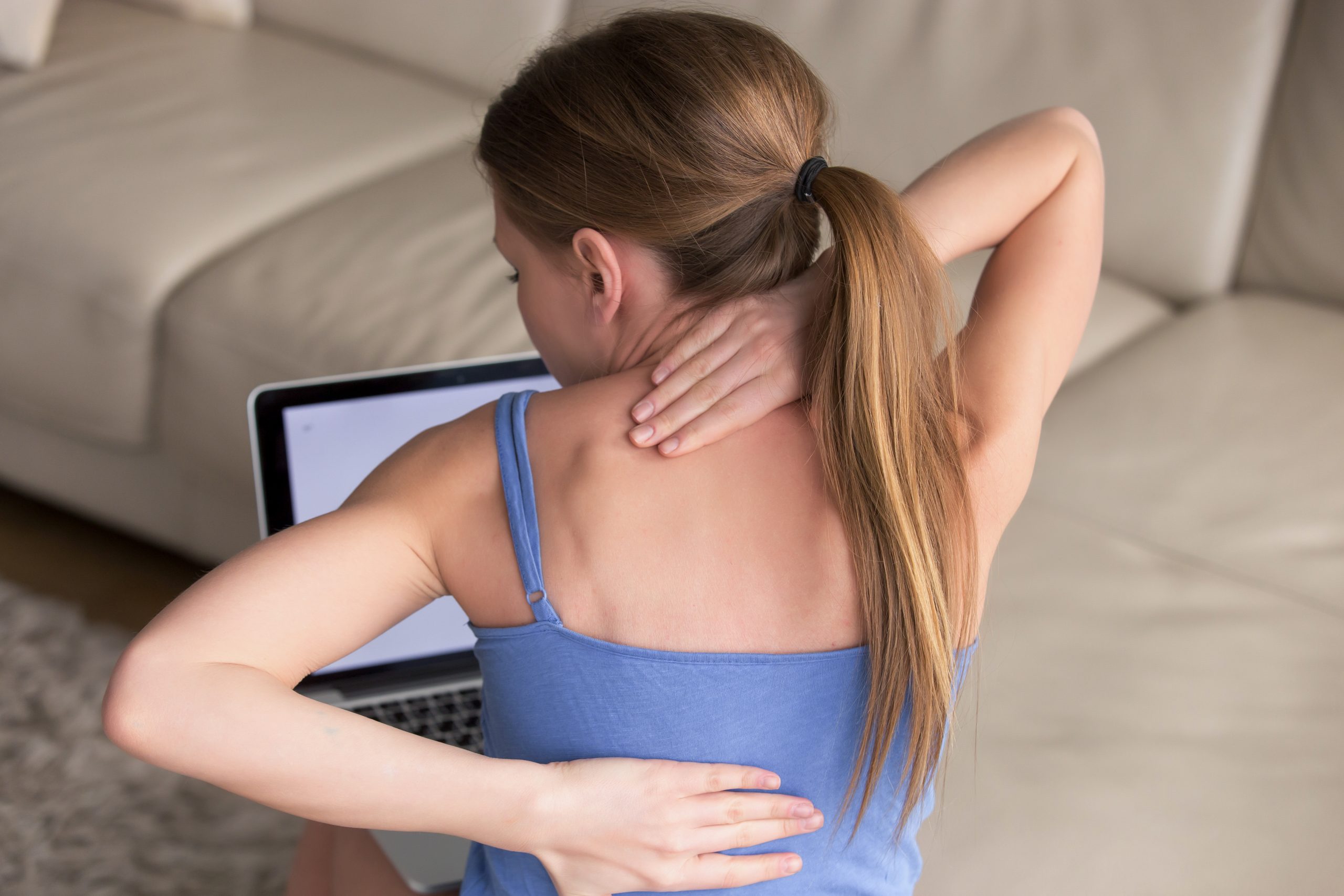
Given the aforementioned information discussed, it is possible to arrive at false conclusions about scoliosis. The parents often complain of the wrong posture of their child, as they are anxious that the wrong position can cause deformation. Scoliosis cannot be caused by everyday actions, such as heavy backpacks, bad posture, or awkward sleeping positions. Moreover, the widespread myth about treatment with massage exists. It is not possible to affect or correct the growth of the vertebra (which is continuously growing) through periodic massages and exercises. There is no scientific evidence about the effectiveness of treatment with special exercises and massages.
Prevention/Timely Detection
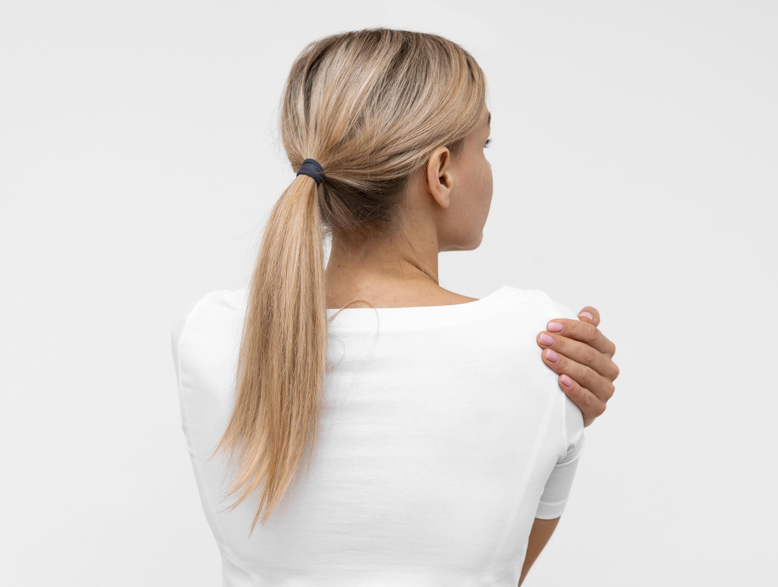
As the cause of the disease is still unknown, it is impossible to prevent. Therefore, the main goal is to detect this pathology as early as possible. The first sign, besides S-shape deformation, is the rotation of the vertebrae along their axes. When the vertebra rotates, it brings structures up on one side and down on the other. This rotation results in the appearance of the so-called rib hump (eminence) on one side of the spine. This is easily noticed when the child stands with legs straight and bends forward (Adam’s test). On one side of the spine, the hump can be easily noticed without special equipment. Parents can do this test themselves at home. Since Adolescent Idiopathic Scoliosis starts during the growth spurt period (around 10 years old), it is important to explain the methodology of Adam’s test to parents so that they can perform this examination at home until the end of the patient’s growth. Referral to a Pediatric orthopedic surgeon is indicated in the cases of signs described above.
Hope this helps!
Best regards,
Dr. Davit Sekoyan, Pediatric Orthopedic Surgeon, “Wigmore” Medical Centre